

Gonadal development of Acropora formosa and Favites abdita in Weizhou Island
-
摘要: 性腺发育是造礁珊瑚有性繁殖的重要过程,珊瑚会在性腺发育成熟后待环境适宜时进行大规模排卵。了解造礁珊瑚的性腺发育过程对预测其排卵时间具有重要的意义,但目前尚未有关于涠洲岛海域造礁珊瑚性腺发育的报道,且该海域造礁珊瑚的排卵时间也不明确。因此,本文于2021年9月至2022年5月,以涠洲岛海域的美丽鹿角珊瑚(Acropora formosa)和秘密角蜂巢珊瑚(Favites abdita)为研究对象,观察其在自然海域的性腺发育过程及排卵时间。结果显示,在自然海域美丽鹿角珊瑚的卵母细胞从9月开始发育,约经9个月发育成熟,精巢从11月开始发育,经2~3个月成熟;秘密角蜂巢珊瑚的卵母细胞在10–11月间开始发育,经7~8个月发育成熟,精巢发育周期为1~2个月;2种珊瑚的配子均在2022年5月份同步成熟,并于2022年5月19–22日(农历四月十九至二十二日)间观察到了室内2种珊瑚的排卵行为,与其在自然海域的排卵时间基本一致。基于上述证据,本文推测涠洲岛海域美丽鹿角珊瑚和秘密角蜂巢珊瑚主要的排卵时间在农历四月十五日前后。本研究为涠洲岛造礁珊瑚的繁殖生物学提供了宝贵的信息,为进一步利用其有性繁殖进行珊瑚礁的生态修复等提供了理论依据。Abstract: Gonadal development is a crucial process for sexual reproduction in scleractinian corals, after the gonad matured, corals will spawn in suitable environments, understanding this process is essential for predicting their spawn time. However, there is no report on the gonadal development cycle and the spawning time of corals in Weizhou Island is currently unknown. From September 2021 to May 2022, our study focused on Acropora formosa and Favites abdita in Weizhou Island to observe their gonadal development process and spawning time. In the wild, the oocytes of A. formosa began to develop in September and matured after nine months, while the testes were observed in November and matured after two to three months. In F. abdita, the onset of oocytes was between in October and November, mature oocytes were observed after seven to eight months, with the development period of the testes was about one to two months. Both corals’ gametes matured in May. In tanks, both corals were observed to spawn between May 19 to 22, 2022 ( April 19 to 22, Chinese lunar calendar), which was consistent with their spawning observed in wild. Based on the results, we postulate that the spawning time of A. formosa and F. abdita in Weizhou Island is around April 15th, Chinese lunar calendar. This study provides valuable information on the reproductive biology of Weizhou Island corals.
-
Key words:
- scleratinian coral /
- spawning time /
- gonad /
- histological analyses /
- sexual reproduction /
- Weizhou Island
-

图 1 美丽鹿角珊瑚的性腺发育过程及排卵行为
a. 第Ⅰ时相卵母细胞(2021年9月28日);b. 第Ⅱ时相卵母细胞(2021年11月18日);c1, c2, c3. 在不同肠系膜中发育的第Ⅲ时相卵母细胞(c1、c2)和第Ⅰ时相精巢(c1、c3)(2022年3月8日);d1, d2. 第Ⅲ时相卵母细胞(d1)和第Ⅱ时相精巢(d2)(2022年4月28日);e1, e2. 第Ⅳ时相卵母细胞(e1)和第Ⅲ时相精巢(e2)(2022年5月8日);f1, f2. 第Ⅳ时相的精巢(f1)、光学显微镜下发育成熟的性腺(f2)(2022年5月18日);g1, g2, g3. 养殖缸(g1)和海区(g3)正在排卵的美丽鹿角珊瑚、排出的卵包(g2)(白色箭头所示为卵包)(2022年5月19日至22日);h. 排卵后的性腺(2022年5月25日);O. 卵母细胞;N. 细胞核;n. 核仁;lv. 脂质泡;cgb. 刺丝囊;sp. 精巢;yg. 卵黄颗粒;sni. 精原细胞;me. 中胶层;sti. 精母细胞;sdi. 精细胞;szoi. 精子;t. 精子尾. 标尺如下:a, b, c3, d2, e2, f1为50 μm;c1, c2, d1, e1为200 μm;f2, g2, h为500 μm
Fig. 1 Gonadal development and spawning of Acropora formosa
a. Stage Ⅰ oocyte (September 28, 2021); b. Stage Ⅱ oocyte (November 18, 2021); c1, c2, c3. Stage Ⅲ oocyte (c1, c2) and Stage Ⅰ spermary (c1, c3) developing in different mesenteries (March 8, 2022); d1, d2. Stage Ⅲ oocyte (d1) and Stage Ⅱ spermary (d2) (April 28, 2022); e1, e2. Stage Ⅳ oocyte (e1) and Stage Ⅲ spermary (e2) (May 8, 2022); f1, f2. Stage Ⅳ spermary (f1), mature gonads (f2) (May 18, 2022); g1, g2, g3. A. formosa were spawning in tank (g1) and in the wild (g3), egg-sperm bundle (g2) (egg-sperm bundles shown by the white arrow) (May 19–22, 2022); h. mesenteries after spawning (May 25, 2022); O. oocyte; N. nucleus; n. nucleolus; lv. lipid vesicle; cgb. cnidoglandular band; sp. spermary; yg. yolk granules; sni. spermatogonia; me. mesoglea; sti. spermatocyte; sdi. spermatid; szoi. spermatozoon; t. sperm tail. a, b, c3, d2, e2, f1: scales = 50 μm; c1, c2, d1, e1: scales = 200 μm; f2, g2, h: scales = 500 μm
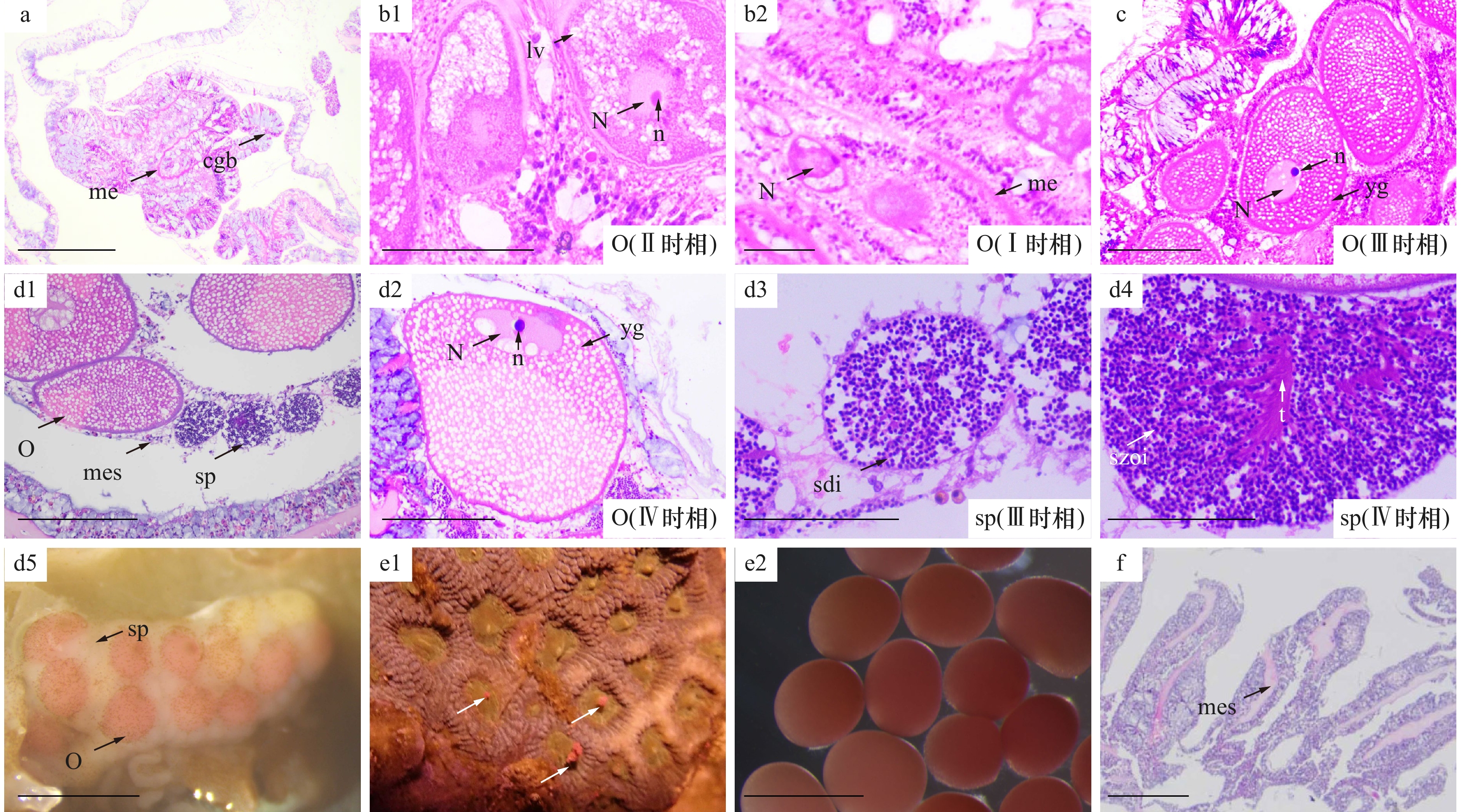
图 2 秘密角蜂巢珊瑚的性腺发育过程及排卵行为
a. 性腺未发育(2021年 9月28日);b1, b2. 第Ⅱ时相卵母细胞(b1)和第Ⅰ时相卵母细胞(b2)(2021年 11月18日);c. 第Ⅲ时相卵母细胞(2022年 3月8日);d1, d2, d3, d4, d5. 在同一肠系膜中发育的配子(d1),第Ⅳ时相卵母细胞(d1、d2),第Ⅲ、Ⅳ时相精巢(d3、d4)及光学显微镜下成熟的性腺(d5)(2022年5月8日);e1, e2. 养殖缸内正在排卵的秘密角蜂巢珊瑚(e1)(白色箭头所示为卵包)及卵母细胞(e2)(2022年 5月22日);f. 排卵后的性腺(2022年5月25日);me. 中胶层;cgb. 刺丝囊;O. 卵母细胞;N. 细胞核;n. 核仁;lv. 脂质泡;yg. 卵黄颗粒;mes. 肠系膜;sp. 精巢;sdi. 精细胞;szoi. 精子;t. 精子尾. 标尺如下:b1, b2, d3, d4为50 μm;a, c, d1, d2为200 μm;d5, e2, f为500 μm
Fig. 2 Gonadal development and spawning of Favites abdita
a. Undeveloped gonad (September 28, 2021); b1.b2. Stage Ⅱ oocyte (b1) and Stage Ⅰ oocyte (b2) (November 18, 2021); c. Stage Ⅲ oocyte (March 8, 2022); d1, d2, d3, d4, d5. gametes developing in the same mesentery (d1), Stage Ⅳ oocyte (d1 , d2), Stage Ⅲ and Ⅳ spermaries (d3, d4) , mature gonad (d5) (May 8, 2022); e1, e2. F. abdita were spawning in tank (e1) (egg-sperm bundles shown by the white arrow) and oocytes (e2) (May 22, 2022); f. mesenteries after spawning (May 25, 2022); me. mesoglea; cgb. cnidoglandular band; O. oocyte; N. nucleus; n. nucleolus; lv. lipid vesicle; yg. yolk granules; mes. mesentery; sp. spermary; sdi. spermatid; szoi. spermatozoon; t. sperm tail. b1, b2, d3, d4: scales = 50 μm; a, c, d1, d2: scales = 200 μm; d5, e2, f: scales = 500 μm
表 1 不同时期造礁珊瑚配子的主要形态特征
Tab. 1 Morphological characteristics of different gonadal stages in scleractinian corals
配子类型 时期 主要形态特征 图示 美丽鹿角珊瑚 秘密角蜂巢珊瑚 卵母细胞 Ⅰ时相 细胞质质密,核质比较高,核膜不清晰 图1a 图2b2 Ⅱ时相 卵母细胞内出现脂质泡,形状不规则 图1b 图2b1 Ⅲ时相 出现卵黄颗粒,细胞核向细胞一端迁移 图1c2、图1d1 图2c Ⅳ时相 细胞间紧密黏连,细胞膜表面不规则凹陷 图1e1 图2d2 精巢 Ⅰ时相 较薄的中胶层包裹着十几个精原细胞 图1c3 − Ⅱ时相 精母细胞排列紧密,中胶层形成较厚的壁 图1d2 − Ⅲ时相 中胶层变薄,精细胞排列疏松,精巢内出现空腔 图1e2 图2d3 Ⅳ时相 精子紧密排列,尾部汇聚于一侧 图1f1 图2d4 注:−表示无数据。  下载: 导出CSV
-
[1] Richmond R H. Reproduction and recruitment in corals: critical links in the persistence of reefs[M]//Birkeland C. Life and Death of Coral Reefs. New York: Chapman & Hall, 1997. [2] Szmant A M. Reproductive ecology of Caribbean reef corals[J]. Coral Reefs, 1986, 5(1): 43−53. doi: 10.1007/BF00302170 [3] Baird A H, Guest J R, Willis B L. Systematic and biogeographical patterns in the reproductive biology of scleractinian corals[J]. Annual Review of Ecology, Evolution, and Systematics, 2009, 40: 551−571. doi: 10.1146/annurev.ecolsys.110308.120220 [4] Loya Y, Sakai K. Bidirectional sex change in mushroom stony corals[J]. Proceedings of the Royal Society B: Biological Sciences, 2008, 275(1649): 2335−2343. doi: 10.1098/rspb.2008.0675 [5] Kersting D K, Casado C, López-Legentil S, et al. Unexpected patterns in the sexual reproduction of the Mediterranean scleractinian coral Cladocora caespitosa[J]. Marine Ecology Progress Series, 2013, 486: 165−171. doi: 10.3354/meps10356 [6] Goffredo S, Dubinsky Z. The Cnidaria, Past, Present and Future: The World of Medusa and Her Sisters[M]. Switzerland: Springer, 2016. [7] Harrison P L, Wallace C C. Reproduction, dispersal and recruitment of scleractinian corals[M]//Dubinsky Z. Ecosystems of the World: Coral Reefs. Amsterdam: Elsevier, 1990: 133−207. [8] Harrison P L. Sexual reproduction of scleractinian corals[M]//Dubinsky Z, Stambler N. Coral Reefs: An Ecosystem in Transition. Dordrecht: Springer, 2011: 59−85. [9] Fadlallah Y H. Sexual reproduction, development and larval biology in scleractinian corals: a review[J]. Coral Reefs, 1983, 2(3): 129−150. doi: 10.1007/BF00336720 [10] Sawall Y, Al-Sofyani A. Biology of Red Sea corals: metabolism, reproduction, acclimatization, and adaptation[M]//Rasul N M A, Stewart I C F. The Red Sea: The Formation, Morphology, Oceanography and Environment of a Young Ocean Basin. Berlin, Heidelberg: Springer, 2015: 487−509. [11] Oliver J, Babcock R C, Harrison P L, et al. Geographic extent of mass coral spawning: clues to ultimate causal factors[C]//Proceedings of the Sixth International Coral Reef Symposium. Townsville: [s.n.], 1988: 803−810. [12] Szmant-Froelich A, Yevich P, Pilson M E Q. Gametogenesis and early development of the temperate coral Astrangia danae (Anthozoa: Scleractinia)[J]. The Biological Bulletin, 1980, 158(2): 257−269. doi: 10.2307/1540935 [13] Glynn P W, Gassman N J, Eakin C M, et al. Reef coral reproduction in the eastern Pacific: Costa Rica, Panama, and Galapagos Islands (Ecuador) I. Pocilloporidae[J]. Marine Biology, 1991, 109(3): 355−368. doi: 10.1007/BF01313501 [14] Gilmour J P, Underwood J N, Howells E J, et al. Biannual spawning and temporal reproductive isolation in Acropora corals[J]. PLoS One, 2016, 11(3): e0150916. doi: 10.1371/journal.pone.0150916 [15] Nozawa Y, Tokeshi M, Nojima S. Reproduction and recruitment of scleractinian corals in a high-latitude coral community, Amakusa, southwestern Japan[J]. Marine Biology, 2006, 149(5): 1047−1058. doi: 10.1007/s00227-006-0285-5 [16] Soto D, Weil E. Sexual reproduction in the Caribbean coral genus Isophyllia (Scleractinia: Mussidae)[J]. PeerJ, 2016, 4: e2665. doi: 10.7717/peerj.2665 [17] Mangubhai S, Harrison P L. Gametogenesis, spawning and fecundity of Platygyra daedalea (Scleractinia) on equatorial reefs in Kenya[J]. Coral Reefs, 2008, 27(1): 117−122. doi: 10.1007/s00338-007-0297-8 [18] 李元超, 黄晖, 董志军, 等. 鹿回头佳丽鹿角珊瑚卵母细胞发育的组织学研究[J]. 热带海洋学报, 2009, 28(1): 56−60. doi: 10.3969/j.issn.1009-5470.2009.01.009Li Yuanchao, Huang Hui, Dong Zhijun, et al. A histological analysis on oocyte development of Acropora pulchra in Sanya of Hainan Island[J]. Journal of Tropical Oceanography, 2009, 28(1): 56−60. doi: 10.3969/j.issn.1009-5470.2009.01.009 [19] 张诗泽, 黄晖, 张浴阳, 等. 鹿回头多孔鹿角珊瑚与丛生盔形珊瑚性腺组织学研究[J]. 生态科学, 2016, 35(1): 41−46.Zhang Shize, Huang Hui, Zhang Yuyang, et al. Histological analyses of the gonad for Acropora millepora and Galaxea fascicularis from Sanya Luhuitou of Hainan Island[J]. Ecological Science, 2016, 35(1): 41−46. [20] 杨小东. 澄黄滨珊瑚、大管孔珊瑚和丛生盔形珊瑚性腺发育与生长规律的研究[D]. 湛江: 广东海洋大学, 2013.Yang Xiaodong. Study of gonad development and growths of Porites lutea, Goniopora djiboutiensis and Galaxea fascicularis[D]. Zhanjiang: Guangdong Ocean University, 2013. [21] 金磊. 盾形陀螺珊瑚和稀杯盔形珊瑚性腺发育与生长规律的研究[D]. 湛江: 广东海洋大学, 2014.Jin Lei. Study of gonad development and growths of Turbinaria peltata and Galaxea astreata[D]. Zhanjiang: Guangdong Ocean University, 2014. [22] Chen C J, Chen W J, Chang C F. Multispecies spawning of scleractinian corals in nonreefal coral communities of northern Taiwan in the northwestern Pacific Ocean[J]. Bulletin of Marine Science, 2021, 97(2): 351−371. doi: 10.5343/bms.2020.0058 [23] 王文欢. 近30年来北部湾涠洲岛造礁石珊瑚群落演变及影响因素[D]. 南宁: 广西大学, 2017.Wang Wenhuan. Evolvement and influential factors of coral community over past three decases in Weizhou Island reef, Beibu Gulf[D]. Nanning: Guangxi University, 2017. [24] Yu Wanjun, Wang Wenhuan, Yu Kefu, et al. Rapid decline of a relatively high latitude coral assemblage at Weizhou Island, northern South China Sea[J]. Biodiversity and Conservation, 2019, 28(14): 3925−3949. doi: 10.1007/s10531-019-01858-w [25] 王欣, 高霆炜, 陈骁, 等. 涠洲岛园艺式珊瑚苗圃的架设与移植[J]. 广西科学, 2017, 24(5): 462−467.Wang Xin, Gao Tingwei, Chen Xiao, et al. The construction and transplantation of coral gardening nursery in Weizhou Island[J]. Guangxi Sciences, 2017, 24(5): 462−467. [26] Wallace C C. Reproduction, recruitment and fragmentation in nine sympatric species of the coral genus Acropora[J]. Marine Biology, 1985, 88(3): 217−233. doi: 10.1007/BF00392585 [27] 于婉君. 涠洲岛珊瑚礁区的底质特征及其对珊瑚分布的影响[D]. 南宁: 广西大学, 2022.Yu Wanjun. Substrate characteristics of the area of Weizhou Island reef and ITS effects on the distribution of corals[D]. Nanning: Guangxi University, 2022. [28] Kojis B L, Quinn N J. Reproductive ecology of two faviid corals (coelenterata: scleractinia)[J]. Marine Ecology Progress Series, 1982, 8(3): 251−255. [29] Maboloc E A, Jamodiong E A, Villanueva R D. Reproductive biology and larval development of the scleractinian corals Favites colemani and F. abdita (Faviidae) in northwestern Philippines[J]. Invertebrate Reproduction & Development, 2016, 60(1): 1−11. [30] Lin C H, Nozawa Y. The influence of seawater temperature on the timing of coral spawning[J]. Coral Reefs, 2023, 42(2): 417−426. doi: 10.1007/s00338-023-02349-9 [31] Shikina S, Chang C F. Sexual reproduction in stony corals and insight into the evolution of oogenesis in Cnidaria[M]//Goffredo S, Dubinsky Z. The Cnidaria, Past, Present and Future: The World of Medusa and Her Sisters. Cham: Springer, 2016: 249−268. [32] Gomez E J, Jamodiong E A, Maboloc E A, et al. Gametogenesis and reproductive pattern of the reef-building coral Acropora millepora in northwestern Philippines[J]. Invertebrate Reproduction & Development, 2018, 62(4): 202−208. [33] Munasik M, Widyatmoko W. Reproduksi karang Acropora aspera di Pulau Panjang, Jawa Tengah: I. Gametogenesis[J]. Indonesian Journal of Marine Sciences, 2004, 9(4): 211−216. [34] Huang Boyin, Thorne P W, Banzon V F, et al. Extended reconstructed sea surface temperature, version 5 (ERSSTv5): upgrades, validations, and intercomparisons[J]. Journal of Climate, 2017, 30(20): 8179−8205. doi: 10.1175/JCLI-D-16-0836.1 [35] Fan T Y, Dai Changfeng. Reproductive plasticity in the reef coral Echinopora lamellosa[J]. Marine Ecology Progress Series, 1999, 190: 297−301. doi: 10.3354/meps190297 [36] Rossi S, Gili J M, Coma R, et al. Temporal variation in protein, carbohydrate, and lipid concentrations in Paramuricea clavata (Anthozoa, Octocorallia): evidence for summer–autumn feeding constraints[J]. Marine Biology, 2006, 149(3): 643−651. doi: 10.1007/s00227-005-0229-5 [37] Baird A H, Guest J R, Edwards A J, et al. An Indo-Pacific coral spawning database[J]. Scientific Data, 2021, 8(1): 35. doi: 10.1038/s41597-020-00793-8 [38] 韦芬, 黄雯, 余克服, 等. 广西涠洲岛黄癣蜂巢珊瑚、肉质扁脑珊瑚的胚胎和幼虫的早期发育[J]. 海洋学报, 2020, 42(4): 87−95.Wei Fen, Huang Wen, Yu Kefu, et al. Embryonic and larval early development of Favia favus and Platygyra carnosus in the Weizhou Island, Guangxi[J]. Haiyang Xuebao, 2020, 42(4): 87−95. [39] Babcock R C, Bull G D, Harrison P L, et al. Synchronous spawnings of 105 scleractinian coral species on the Great Barrier Reef[J]. Marine Biology, 1986, 90(3): 379−394. doi: 10.1007/BF00428562 [40] Shlesinger T, Loya Y. Breakdown in spawning synchrony: a silent threat to coral persistence[J]. Science, 2019, 365(6457): 1002−1007. doi: 10.1126/science.aax0110 [41] Guest J R, Baird A H, Goh B P L, et al. Seasonal reproduction in equatorial reef corals[J]. Invertebrate Reproduction & Development, 2005, 48(1/3): 207−218. [42] Fogarty N D, Marhaver K L. Coral spawning, unsynchronized[J]. Science, 2019, 365(6457): 987−988. doi: 10.1126/science.aay7457 [43] Keith S A, Maynard J A, Edwards A J, et al. Coral mass spawning predicted by rapid seasonal rise in ocean temperature[J]. Proceedings of the Royal Society B: Biological Sciences, 2016, 283(1830): 20160011. doi: 10.1098/rspb.2016.0011 [44] 黄洁英, 黄晖, 张浴阳, 等. 膨胀蔷薇珊瑚与壮实鹿角珊瑚的胚胎和幼虫发育[J]. 热带海洋学报, 2011, 30(2): 67−73. doi: 10.3969/j.issn.1009-5470.2011.02.010Huang Jieying, Huang Hui, Zhang Yuyang, et al. Embryonic and larval development of Montipora turgescens and Acropora robusta[J]. Journal of Tropical Oceanography, 2011, 30(2): 67−73. doi: 10.3969/j.issn.1009-5470.2011.02.010 [45] Lin C H, Nozawa Y. Variability of spawning time (lunar day) in Acropora versus merulinid corals: a 7-yr record of in situ coral spawning in Taiwan[J]. Coral Reefs, 2017, 36(4): 1269−1278. doi: 10.1007/s00338-017-1622-5 [46] Randall C J, Negri A P, Quigley K M, et al. Sexual production of corals for reef restoration in the Anthropocene[J]. Marine Ecology Progress Series, 2020, 635: 203−232. doi: 10.3354/meps13206 [47] Komoto H, Lin C H, Nozawa Y, et al. An external coincidence model for the lunar cycle reveals circadian phase-dependent moonlight effects on coral spawning[J]. Journal of Biological Rhythms, 2023, 38(2): 148−158. doi: 10.1177/07487304221135916 [48] Shoguchi E, Tanaka M, Shinzato C, et al. A genome-wide survey of photoreceptor and circadian genes in the coral, Acropora digitifera[J]. Gene, 2013, 515(2): 426−431. doi: 10.1016/j.gene.2012.12.038 [49] Lin C H, Takahashi S, Mulla A J, et al. Moonrise timing is key for synchronized spawning in coral Dipsastraea speciosa[J]. Proceedings of the National Academy of Sciences of the United States of America, 2021, 118(34): e2101985118. [50] Kaniewska P, Alon S, Karako-Lampert S, et al. Signaling cascades and the importance of moonlight in coral broadcast mass spawning[J]. eLife, 2015, 4: e09991. doi: 10.7554/eLife.09991 [51] Shima J S, Osenberg C W, Alonzo S H, et al. How moonlight shapes environments, life histories, and ecological interactions on coral reefs[J]. Emerging Topics in Life Sciences, 2022, 6(1): 45−56. doi: 10.1042/ETLS20210237 [52] Wolstenholme J, Nozawa Y, Byrne M, et al. Timing of mass spawning in corals: potential influence of the coincidence of lunar factors and associated changes in atmospheric pressure from northern and southern hemisphere case studies[J]. Invertebrate Reproduction & Development, 2018, 62(2): 98−108. -

计量
- 文章访问数: 379
- HTML全文浏览量: 152
- PDF下载量: 38
- 被引次数: 0
